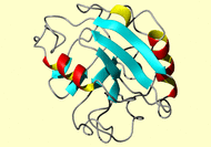
NMR Restraints Grid

 |
NMR Restraints Grid |
 |
Result table
| image | mrblock_id | pdb_id | bmrb_id | cing | stage | program | type |
|
|
2799 |
1clh |
4037 | cing | 1-original | MR format | comment |
*HEADER    ISOMERASE(PEPTIDYL-PROLYL CIS-TRANS)    20-DEC-93   1CLH
*COMPND    CYCLOPHILIN (NMR, 12 STRUCTURES)
*SOURCE    (ESCHERICHIA COLI)
*AUTHOR    R.T.CLUBB,G.WAGNER
*REVDAT   1   31-MAY-94 1CLH    0
##############################################################################

SUPPLEMENTARY MATERIAL

  Experimental NMR restraints used to determine the three-dimensional solution
structure of periplasmic Cyclophilin from E. coli. The structures are based
on 1458 interproton distance restraints derived from NOE measurements; 108
hydrogen-bonding distance restraints for 54 hydrogen-bonds identified on the
basis of the NOE and amide proton exchange data, as well as initial structure
calculations; and 101 phi and 53 chi1 torsion angle restraints derived from
coupling constants and NOE data.

All the coordinates are included as a separate file: ecyp_brookhaven.pdb

References

1.	Clubb, R. T., Ferguson, S. B., Walsh, C. T., and Wagner, G. (1994)
	Biochemistry, in press

##############################################################################

NOE RESTRAINTS

1458	Total
483 	(|i-j|) =  1
149	(|i-j|) =< 4
574	(|i-j|) > 4
252	Intraresidual

("-1.0" indicates a lower bound equal to the sum of the van der Waals radii)

##############################################################################
				      Bounds
Residue	Atom	Residue	Atom	Lower(A)  Upper(A)
##############################################################################
Contact the webmaster for help, if required. Sunday, April 28, 2024 3:38:17 PM GMT (wattos1)