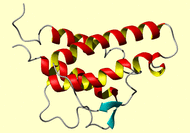
NMR Restraints Grid

 |
NMR Restraints Grid |
 |
Result table
| image | mrblock_id | pdb_id | bmrb_id | cing | in_dress | stage | program | type |
|
|
2021 |
1bbn |
4094 | cing | dress | 1-original | MR format | comment |
*HEADER CYTOKINE 01-MAY-92 1BBN *COMPND INTERLEUKIN 4 (NMR, MINIMIZED AVERAGE STRUCTURE) *SOURCE HUMAN (HOMO SAPIENS) RECOMBINANT FORM EXPRESSED IN YEAST *SOURCE 2 (SACCHAROMYCES CEREVISIAE) *AUTHOR G.M.CLORE,B.POWERS,D.S.GARRETT,A.M.GRONENBORN *REVDAT 1 31-OCT-93 1BBN 0 REMARK Experimental NMR restraints used for the three-dimensional structure REMARK determination of recombinant human interleukin-4 REMARK REMARK REMARK Authors: G.M. Clore, B. Powers, D.S. Garrett and A.M. Gronenborn REMARK REMARK References REMARK REMARK 1. R. Powers, D.S. Garrett, C.J. March, E.A. Frieden, A.M. Gronenborn REMARK and G.M. Clore (1992) Three dimensional solution structure of REMARK human interleukin-4 by multidimensional heteronuclear magnetic REMARK resonance spectroscopy. Science in press REMARK REMARK 2. R. Powers, D.S. Garrett, C.J. March, E.A. Frieden, A.M. Gronenborn REMARK and G.M. Clore (1992) 1H, 15N, 13C and 13CO assignments of human REMARK interleukin-4 using three-dimensional double- and triple-resonance REMARK hetronuclear magnetic resonance spectroscopy. Biochemistry 31, issue REMARK 18, in press REMARK REMARK 3. D.S. Garrett, R. Powers, C.J. March, E.A. Frieden, G.M. Clore REMARK and A.M. Gronenborn (1992) Determination of the secondary structure REMARK and folding topology of human interleukin-4 using three-dimensional REMARK heteronuclear magnetic resonance spectroscopy. Biochemistry 31, REMARK issue 18, in press REMARK REMARK All the coordinates REMARK are included here as a separate file: il4_brookhaven.pdb REMARK REMARK The numbering scheme in this structure includes the four-residue REMARK sequence Glu-Ala-Glu-Ala at the N-terminus of the recombinant REMARK protein which is not part of the natural human IL-4; the natural REMARK IL-4 sequence therefore starts at residue 5. REMARK REMARK Details of the structure determination and all structural REMARK statistics are given in ref. 1 (i.e. agreement with experimental REMARK restraints, deviations from ideality for bond lengths, angles, REMARK planes and chirality, non-bonded contacts, atomic rms differences REMARK between the calculated structures). REMARK The structures are based on 823 interproton distance restraints REMARK derived from NOE measurements; 98 hydrogen-bonding distance REMARK restraints for 49 hydrogen-bonds identified on the basis of the REMARK NOE and amide proton exchange data, as well as the initial structure REMARK calculations; and 101 phi and 82 psi backbone torsion angle REMARK restraints derived from oupling constants, NOE data, and 13C REMARK secondary chemical shifts. REMARK REMARK The method used to determine the structures REMARK is the hybrid metric matrix distance geometry-dynamical simulated REMARK annealing method [Nilges, M., Clore, G.M. & Gronenborn, A.M. REMARK FEBS Lett. 229, 317-324 (1988)]. REMARK REMARK REMARK REMARK The NOE restraints are given in (A) and the torsion angle restraints REMARK in (B). REMARK REMARK A. NOE interproton distance restraints The restraints are represented by square-well potentials with the upper (u) and lower (l) limits given by u=i+k and l=i-j where the numbers are entered in the order i,j,k. [Clore et al. (1986) EMBO J. 5, 2729-2735] The NOEs are classified into three distance ranges corresponding to strong, medium and weak NOEs. These are 1.8-2.7 A, 1.8-3.3 A and 1.8-5.0 A, respectively. Appropriate corrections to the upper limits for distances involving methyl, methylene and Tyr and Phe aromatic ring protons, to account for centre averaging, are carried out as described by Wuthrich et al. [J. Mol. Biol. 169, 949-961 (1983)]. In addition, an extra 0.5 A is added to the upper limits of distances involving methyl protons [Clore et al. (1983) Biochemistry 26, 8012-8023; Wagner et al. (1987) J. Mol. Biol. 196, 611-640]. The atom notation follows standard PDB format. The # indicates a single wild card, and the * a full wild card. e.g. For Leu, HD* representes all the methyl protons; for a normal methylene beta proton, HB# represents the two protons. In these cases, the distances are calculated as centreaverages. Note that the hard sphere van der Waals repulsion term ensures that the minimum lower limit for all distances is the sum of the relevant hard sphere atom radii.
Contact the webmaster for help, if required. Thursday, May 2, 2024 10:57:44 AM GMT (wattos1)